Post-Arrest Prognostication

While I want to focus this blog on things relevant to practice in the Emergency Department, I have an academic interest (and maybe a career interest long-term) in critical care. I also feel that cardiac arrest is a particular area in critical care should be something that EPs are expert in — it’s also an area in which there is considerable nihilism which may lead in sub-optimal patient care, or early withdrawal of efforts before such withdrawal is justifiable.

What do I mean by nihilism? I mean that we in the ED rarely see good outcomes in out-of-hospital cardiac arrest (OOHCA) patients (and when we do, they’re often comatose and whisked away to the ICU, which means that even if they *do* have a good clinical outcome we do not see it happen and rarely even hear about it), and this leads to a sentiment that any cardiac arrest patient is bound for either death or a meaningless life due to neurologic injury.

Everyone in in the department, from patient care assistants and techs and medical students to the attendings, puts a lot of energy and effort into running codes and trying to resuscitate these patients. People care a lot and do some of their best work in these stressful contexts. But at the same time, I sometimes wonder whether we would focus more on improving our process and quality of care– and perhaps thereby do even better– if we had a better sense that our interventions translated into patients who could again be alive and well because of them. This sense is difficult to come by if many of the patients that you successfully attain ROSC on have features that many people associate with a very low likelihood of meaningful recovery.

This pair of recent review articles focused on prognostication in post-cardiac arrest patients — findings on clinical exam, imaging, and other methods to try to suss out who will go onto do well and who is unlikely to ever regain meaningful neurologic function. As ICU bed availability dwindles and the incidence of cardiac arrest and survival thereof continues to increase, this will be of increasing relevance to ED docs, intensivists, and those working with these patients.

So what is a “good outcome”? As the article says, “Experts in coma prognositication defined outcome by cerebral performance categories (CPCs; CPC 1 back to baseline, CPC 2 moderate impairment, CPC 3 severe impairment, CPC 4 vegetative or comatose, CPC 5: dead).” They bifurcate these into either a good (CPC 1 or 2) or poor (CPC 3-5) outcome. Obviously the difference between “moderate” and “severe” impairment is somewhat subjective, but there are additional tools used to help with this distinction.

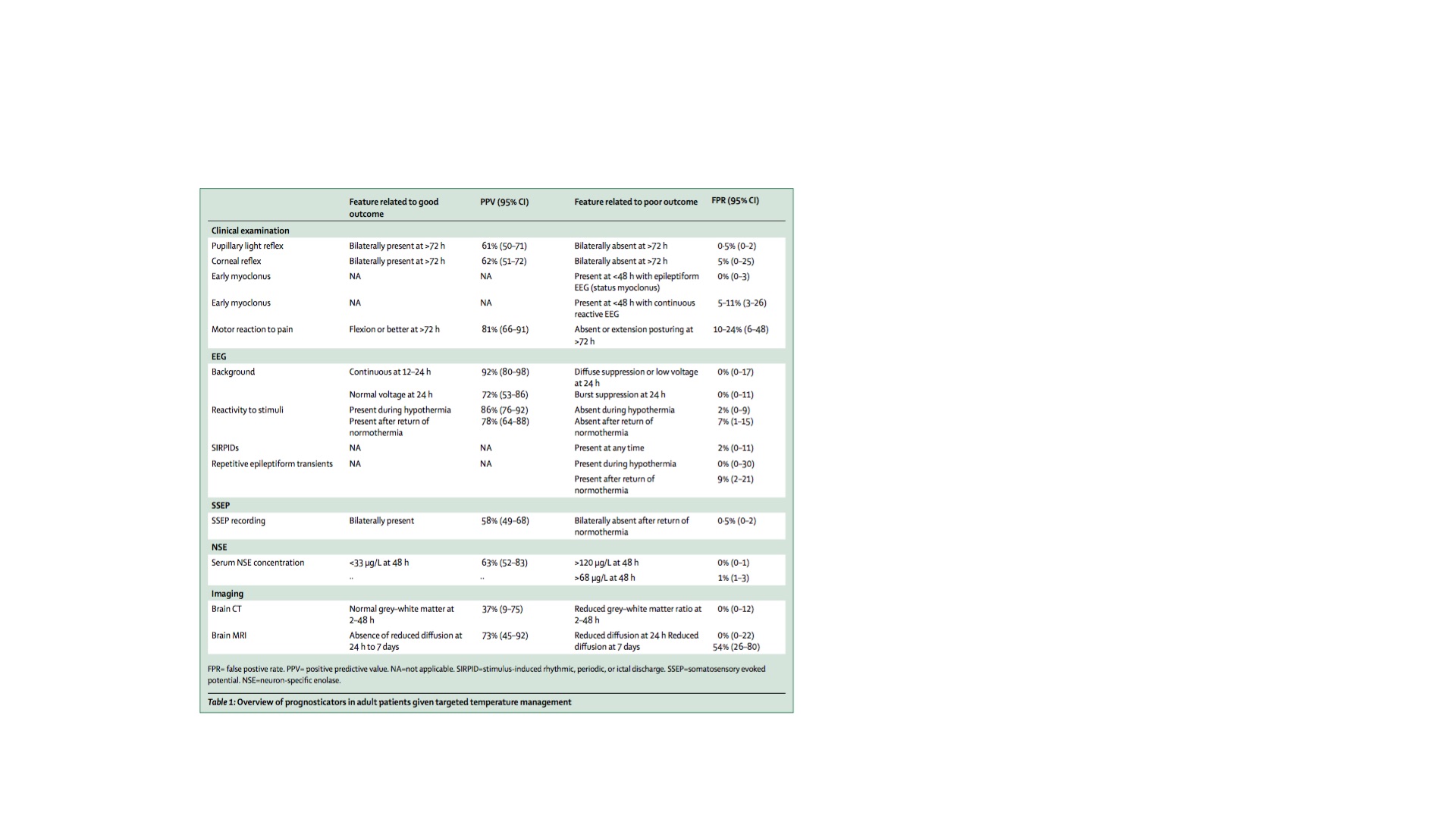
The old standard was clinical assessment of brainstem reflexes, the response to pain, and the absence or presence of myoclonus during the first 72 hours post-arrest. In the TTM era, this becomes trickier because temperature management and the required sedation can alter these features, though the bedside exam still has significant prognostic significance. Absence of pupillary reflexes at 72 hours is the best bedside predictor of a bad outcome, with a false positive rate (FPR) of only 0.5% — presence of pupillary reflexes however, does not confer a good outcome, given that it only has a PPV of 61% (95% CI 50-71).

What about earlier? In the first 24 hours post-arrest, particularly in hypothermic patients, ~ 8% of patients without pupillary reflexes will go on to have a good recovery — so don’t count them out. In terms of corneal reflexes, the reliability is less than that of pupillary reflexes but their absence still correlates with a poor prognosis, with an FPR of 5%.

Motor response is the most affected by sedatives, opiates, and neuromuscular blockade — all common in patients undergoing TTM, and absent or extensor responses to painful stimuli at 72 hours had a FPR of 24%. To reliably utilize this for prognostication, you need exclusion of residual effects of sedation, which can be extended beyond when the drips are simply turned off secondary to the effects of TTM and also the effects of reduced clearance due to shock liver, renal dysfunction, or both.

In terms of myoclonus, which is classically associated with poor outcomes, ~ 9% of patients with myoclonus may survive, according to the data presented here. As the article states, myoclonus is somewhat of a nebulously defined entity — “Not all so-called twitches have the same prognostic implication, rather their usefulness in predicting prognosis depends on semiology, duration, and associated EEG findings.”

I’ll skip EEG and ERPs because this is already too long, but suffice to say they’re useful after hypothermia and for ruling out sub-clinical status epilepticus, which is something we really want to avoid happening in our post-arrest patients, but is very common. More to come on this, which I feel is of particular relevance to us in the ED. Same goes for biomarkers such as neuron specific enolase and Serum S-100B, which can both be measured and trended as the “troponins of the brain”, so to speak.

In terms of imaging — CT scan of the head is recommended in patients in whom there is not another obvious cause of cardiac arrest, to evaluate for a bleed or ischemic stroke. Evaluation of gray:white ratios can predict poor outcomes, but is less reliable than clinical exam and EEG, and this is true for MRI as well, though again MRI does not add very much prognostic capability beyond what can be achieved with bedside tests and the logistics and cost associated with MRI scans of every comatose survivor of cardiac arrest make this somewhat limited in utility.
PPV for Neuro Findings

So what’s the takeaway from all this? Basically, reliable prognostication after cardiac arrest is hard, but at the same time, it isn’t– don’t do it right away, and if you do, it shouldn’t necessarily be based on your bedside neurologic exam. There are tools that can give us useful information, but rarely certainty, to guide conversations with family. And the reality is that none of them are accurate enough inside the first 48-72 hours, especially in patients who are being cooled. There is a very powerful desire to be able to give families hope, or to caution against hope in a way that changes outcomes before they’ve happened — in my very early-in-development opinion, all you can really tell them is something I heard one of my mentors say to families whose children were in the Pediatric ICU: “Prepare for the worst, and hope for the best.”

I also take away from this that nihilism is an un-useful form of prognsotication in these patients — I have seen patients myself who had unreactive pupils or myoclonic jerks, who went onto walk out of the hospital, fairly neurologically intact. This is even more true if the arrest was witnessed, was a shockable rhythm such as VT or VF, and if the patient received high-quality chest compressions and early defibrillation, preventing lengthy low/no-flow states to the brain.

The message not to take away from this post that I believe in any sense that there is no ability to meaningfully make predictions about the likely outcome of cardiac arrest patients, whether or not you’ve gotten ROSC — there are many other variables not considered in the above article  that predict do reliably predict outcomes such as comorbidities, age of the patient, how long they were down for, the initial rhythm, and an often-overlooked variable in the literature (because it’s tough to quantify): consideration of their quality of life before they suffered a cardiac arrest. I also think that the pragmatic realities of cardiac arrest care — an emotionally charged event where patients are often teetering along a line between life and death, and where decisions have real and immediate impacts on that outcome– may require a sense of somewhat-morbid realism when the outcomes are often so dismal. I just hope that when people are making decisions about termination of efforts (or withdrawal of care post-ROSC) they’re considering all of these things and more, beyond just what their clinical gestalt is.

More to come, I’m sure — I’m especially interested in what happens moving forward in terms of biomarkers, cerebral oximetry, and near-infrared brain imaging to try to determine cerebral oxygenation and metabolism without having to move patients out of the ICU.

References

Rossetti AO1, Rabinstein AA2, Oddo M3. Neurological prognostication of outcome in patients in coma after cardiac arrest. Lancet Neurol. 2016 Mar 23. PMID: 27017468. [PubMed] [Read by QxMD]
Sivaraju A1, Gilmore EJ, Wira CR, Stevens A, Rampal N, Moeller JJ, Greer DM, Hirsch LJ, Gaspard N. Prognostication of post-cardiac arrest coma: early clinical and electroencephalographic predictors of outcome. Intensive Care Med. 2015 Jul;41(7):1264-72. PMID: 25940963. [PubMed] [Read by QxMD]

Pulmonary Embolism in Pregnancy

The diagnosis of pulmonary embolism in pregnant patients is one made difficult by many factors, including a normal elevation in serum d-dimer levels (see below) as well as the additional concern regarding exposure of a developing fetus to the high levels of radiation and contrast associated with CT pulmonary angiography. It is well-known that exogenous estrogen is a risk factor for thromboembolic disease, and while it seems from the data discussed below that pregnancy is not as scarily-high-risk for PE as we might think, we certainly know that pregnancy is a time when homones are running high Add to this the fact that in pregnancy, women are both tachypnic and tachycardic due to normal changes in cardiovascular and respiratory physiology — making a clinical diagnosis that much more difficult.

In these sequentially-published review articles by the PE guru Jeff Kline et al., the authors review the diagnostic dilemma presented by these patients and present the following algorithm:

Microsoft Word - jem_10231_JEM10231.edt

Note the inclusion of the trimester-stratified quantitative d-dimer for patients without a high pretest probability who are PERC negative — this goes against the conventional wisdom that the d-dimer is a worthless test in pregnant women due to the normal elevation found intrapartum. Similar to the way we have begun “age-adjusting” the threshold value of the quantitative d-dimer in non-pregnant patients, they propose that the threshold be “adjusted according to the trimester of pregnancy, as follows: first trimester, 750 ng/mL; second trimester, 1000 ng/mL; third trimester, 1250 ng/mL (assuming a standard cutoff of 500 ng/mL). If the patient has a non-high-pretest probability, has no high-risk features, is PERC negative, and the bilateral ultrasound is negative, and the D-dimer is below the trimester-adjusted values, PE can be ruled out to a reasonable degree of medical certainty.”

They acknowledge the limitations of this approach, including that it hasn’t been prospectively validated, and they do not present any data showing its performance as they’ve been using it, but in cases like this expert opinion is the best we have (so far). He discussed this approach on an episode of ER Cast, and explains it a little bit more in terms of the integration into clinical practice, as well as the role that gestalt can play in risk stratification. 

What I found interesting about this was the idea that the post-partum period is the most risky period of time for women in terms of pulmonary embolism — this echoes what we know about cardiovascular disease in the post-partum period, i.e. when women are autotransfused and their cardiopulmonary physiology is rapidly and massively altered, this presents the highest risk in terms of women with heart failure, valvular abnormalities, or disease entities like peripartum cardiomyopathy. According to the data presented by Kline et al, while the risk increases throughout a pregnancy, 70% of all peripartum PEs occur post partum, and the risk during pregnancy is low (OR 0.4-0.8, depending on trimester) — though, as the authors note, this may not actually reflect that pregnancy is protective against PE but instead suggest that we overtest women for pulmonary embolism during pregnancy, perhaps because of the clinical changes described above. The also cite a large meta-analysis of 23 epidemiologic studies that found PE occuring in only 3 of 10,000 pregnancies.

Another thing that stood out to me while reviewing this article was that for a patient to PERC out of these algorithms, their vital signs must be normal throughout their entire ED stay — normalization of vital signs during an ED visit does not lower the risk of PE, as specifically stated by the authors.


References

Kovac M1, Mikovic Z, Rakicevic L, Srzentic S, Mandic V, Djordjevic V, Radojkovic D, Elezovic I. The use of D-dimer with new cutoff can be useful in diagnosis of venous thromboembolism in pregnancy. Eur J Obstet Gynecol Reprod Biol. 2010 Jan;148(1):27-30. PMID: 19804940. [PubMed] [Read by QxMD]
Kline JA1, Williams GW, Hernandez-Nino J. D-dimer concentrations in normal pregnancy: new diagnostic thresholds are needed. Clin Chem. 2005 May;51(5):825-9. PMID: 15764641. [PubMed] [Read by QxMD]

Complex Febrile Seizure & The Utility of Doing Something(s)

A complex febrile seizure — AKA one occurring with focality, duration > 15 minutes or recurrence within 24 hours, or associated with persistent AMS or post-ictal state — demands a greater amount of testing than a simple one. But to what extent? Which children benefit from neuroimaging, lumbar puncture, EEG testing, and which of these children go onto either have a bad outcome or have something diagnosed on one of those tests?

A group of authors studied 526 patients presenting with their first complex febrile seizure. In two separate papers, 64% received an LP and 50% received emergency head imaging. Of these, 3 patients (0.9%) were found to have acute bacterial meningitis — two of these grew out Strep pneumoniae by CSF culture. Among those with Strep pneumo in the CSF, one was non responsive at presentation and the other had a bulging fontanelle and apnea — the third child was well-appearing at presentation, and had a culture that grew out Strep Pneumo from the blood but the LP was unsuccessful. None of the patients who did not undergo lumbar puncture returned to the hospital with a ABM presentation.

In terms of imaging, 4 of the 526 patients had significant finding — two of these patients had ICH, one had ADEM (acute disseminated encephalomyelitis), and one had focal cerebral edema. Of these patients, 3/4 had obvious findings — nystagmus, emesis, AMS, hemiparesis, and bruising suggestive of NAT.

A second, more recent study performed in a Californian emergency department reported outcomes in 193 patients presenting with new-onset CFS, of which 136 received LP (showing the significant variability that exists between practice environments in terms of this practice). Of these, 14 had CSF pleocytosis, and one (0.5%) went on to be diagnosed with ABM. In a subset of these patients who had a second brief febrile seizure within 24 hours and who received LPs, none were found to have ABM or other serious neurologic disease. Again, this supports the suggestion that in patients without other concerning findings on exam, LP may be deferred — it also suggests (though this is a small patient series) that more than one seizure occurring within 24 hours may be protective in terms of risk stratification for ABM or other serious neurologic illnesses.

Takeaway? Tough to say. It seems that the majority of patients presenting with a complex febrile seizure without “obvious” (always easy to write in retrospect) signs of intra-axial badness go on to do very well, or at least go onto have normal findings on LPs and emergent head imaging. This seems to support the idea that LP and neuroimaging should be selectively added to the workup of a complex febrile seizure, rather than be thought of as necessarily indicated in this patient cohort. That said, guidelines are yet to be published by any leading groups such as ACEP or the AAP in terms of workup for complex febrile seizure, so guidance and support for a “standard of care” is yet to exist — tread carefully, and document thoroughly.

References

Teng D1, Dayan P, Tyler S, Hauser WA, Chan S, Leary L, Hesdorffer D. Risk of intracranial pathologic conditions requiring emergency intervention after a first complex febrile seizure episode among children. Pediatrics. 2006 Feb;117(2):304-8. PMID: 16452347. [PubMed] [Read by QxMD]
Kimia AA1, Ben-Joseph E, Prabhu S, Rudloe T, Capraro A, Sarco D, Hummel D, Harper M. Yield of emergent neuroimaging among children presenting with a first complex febrile seizure. Pediatr Emerg Care. 2012 Apr;28(4):316-21. PMID: 22453723. [PubMed] [Read by QxMD]

More Low-Risk Chest Pain!

In this article published in JAMA Internal Medicine in July of last year, a group of emergency physicians reviewed 11,230 records of patients hospitalized for chest pain with 2 negative troponin tests, nonconcerning initial ED vital signs, and nonischemic, interpretable electrocardiographic findings to determine the incidence of patient-centered adverse events in the short term.

What is interesting and unique about this study is the shift from using MACE (which, as I have discussed before, includes somewhat-nebulously-patient-centered bad outcomes such as need for cardiac revascularization — this is an intervention, not a harm that occurred to a patient due to a lack of intervention) from using their more  “clinically relevant adverse cardiac events” (of course requiring a new catchy acronym, CRACE): (1) life-threatening arrhythmia (ventricular fibrillation, sustained ventricular tachycardia requiring treatment, symptomatic bradycardia or bradyasystole requiring emergent intervention, and any tachydysrhythmia treated with cardioversion); (2) inpatient STEMI; (3) cardiac or respiratory arrest; and (4) death.

Another unique aspect of this study was the enrollment of patients who were sick who met their criteria discussed above– many other studies only considered “low risk patients” to be those without significant comorbidities or CV disease histories (e.g. history of CABG, multiple stents, diabetes, hypertension, etc) . They did exclude patients with LBBB or pacemaker rhythms on EKGs, which would have made identification of ischemia perhaps more difficult.

What did they find? Only four patients out of 7266 meeting the above criteria went on to have any of the primary endpoints. Of these, two were non-cardiac and two were possibly iatrogenic. This is a rate of 0.06% (95% CI 0.02-0.14%), which is much lower than many people would likely guess, and can help inform the discussion we can have with patients when arriving at a disposition. If I am practicing in a community such as the authors’, where short-term follow up with a cardiologist can be arranged, and a patient is reliable, I feel that this data can help me feel more comfortable discharging them with that plan rather than admitting to the hospital, if the patient is comfortable with this.

As Ryan Radecki wrote, the applicability of this hinges on tightly integrated follow up, and we cannot practice “catch and release” medicine. This is also only one data set, and requires prospective validation, and we need to acknowledge that this is not a zero-miss strategy (just like any strategy). That said, there are many potential downsides associated with admission, from costs and downstream sequelae of unnecessary invasive testing to iatrogenic harms, and this study will help better inform our conversation with patients about all of these issues.

References

Weinstock MB1, Weingart S2, Orth F3, VanFossen D4, Kaide C5, Anderson J6, Newman DH7. Risk for Clinically Relevant Adverse Cardiac Events in Patients With Chest Pain at Hospital Admission. JAMA Intern Med. 2015 Jul;175(7):1207-12. PMID: 25985100. [PubMed] [Read by QxMD]

Age-Adjusted D-Dimer

Pulmonary embolism is a commonly-investigated diagnosis in the world of emergency department risk stratification — the presentation of these patients is varied, the ultimate impact on patients of the disease entity itself is questionable when it comes to the less sick end of the spectrum, and the tools we have for diagnosis are associated with significant amounts of radiation and contrast. However, in a practice environment with a low tolerance for missed diagnoses (however questionable the risk:benefit balance of the intervention that would have been performed), we continue to strive to balance the risks and costs of diagnostic testing with the very real risk of progressive disease.

The D-Dimer level is a test used in patients with a low to moderate pretest probability of DVT or PE (and possibly aortic dissection?) — if negative, it will virtually rule out PE, and can help you avoid further testing with CT pulmonary angiography. If positive, further testing is required. So why do emergency physicians hate the D-Dimer? Because while elevation in D-Dimer levels is sensitive for pulmonary embolism or DVT, it is not specific — particularly with cutoff levels of ~ 500 ng/dL, which is the conventional cutoff for a positive test. Elevated D-Dimer levels occur for a multitude of reasons, including liver disease, inflammation, malignancy, trauma, pregnancy, and– most complicating of all– advanced age.

The first of the studies I read this weekend, the ADJUST-PE study, a group of authors had previously retrospectively derived and valid the value of a progressive D-Dimer cutoff adjusted to age in 1712 patients — the optimal age-adjusted cutoff was defined as patient’s age multiplied by 10 in patients 50 years or older. The ADJUST-PE study represented their attempt to prospectively validate the adjustment and to assess its impact on patients in real life. In this multi-center study which enrolled 3324 patients, the age adjusted D-Dimer cut off did very well — only one patient who had a D-Dimer between 500 ng/dL and their age-adjusted cutoff (in other words, someone who would have gotten scanned if they weren’t using the new tool) was found at three month follow up to have a PE, and this was non-fatal. The age adjusted level allowed for safe discharge of patients that might otherwise have been exposed to the costs/potential harms associated with CTPA or treatment of non-hemodynamically significant emboli.

The second study takes the same approach and retrospectively applies the cutoff to 31,094 suspected pulmonary embolism patients presenting to an emergency department in the community. They report data for all ED visits for Kaiser Permanente Southern California members older than 50 years, from 2008 to 2013, who received a D-dimer test after presenting with a chief complaint related to possible PE such as chest pain or dyspnea (due to their focus on PE rather than DVT). The authors excluded patients who underwent ultrasound imaging for DVT for the same reason. What they found was a sensitivity of 92.9% and a specificity of 63.9% for the age-adjusted D-Dimer threshold applied to this population — this compares to 98.0% and 54.4% for the traditional threshold of 500 ng/dL. This is not unsurprising — what I thought was interesting about the second paper was its expansion of the discussion of this testing strategy to include estimates for other harms beyond symptomatic PE that might be missed — specifically, they discuss the incidence of contrast-induced nephropathy, and how changes in testing strategies translate into potential benefits there that may outweigh the harms done by missing clots. These are statistical models, and need to be taken with a grain of salt, but they predict that  “using an age-adjusted D-dimer threshold would miss or delay diagnosis of 26 more pulmonary embolisms than the current standard, but it would prevent 322 cases of contrast- induced nephropathy, 29 cases of severe renal failure, and 19 deaths related to contrast-induced nephropathy in this sample.”

So what will I do with this information? Probably try for better shared decision making and try to avoid CTPA in patients with D-Dimers below the age-adjusted cutoff. I think sharing these numbers with our patients in a comprehensible way, and talking to them about the potential harms associated with testing is the best way forward– this will require further work in terms of identifying the best way to communicate these risks and odds to patients, and as always, trying to balance advocacy for patients, and our ultimate goal of keeping them safe, alive and functional, with the fear of missing a diagnosis or sending someone home with a nebulous non-diagnosis and the possibility of clinical deterioration.

References

Righini M1, Van Es J2, Den Exter PL3, Roy PM4, Verschuren F5, Ghuysen A6, Rutschmann OT7, Sanchez O8, Jaffrelot M9, Trinh-Duc A10, Le Gall C11, Moustafa F12, Principe A13, Van Houten AA14, Ten Wolde M15, Douma RA2, Hazelaar G16, Erkens PM17, Van Kralingen KW18, Grootenboers MJ19, Durian MF20, Cheung YW15, Meyer G8, Bounameaux H1, Huisman MV3, Kamphuisen PW21, Le Gal G22. Age-adjusted D-dimer cutoff levels to rule out pulmonary embolism: the ADJUST-PE study. JAMA. 2014 Mar 19;311(11):1117-24. PMID: 24643601. [PubMed] [Read by QxMD]

Duration of symptoms of respiratory tract infections in children

From the BMJ, we have a very interesting systematic review evaluating the duration of symptoms in children seen in the ED (or A&E, if you will) for fairly minor complaints: otitis media, acute cough, sore throat, and common cold. In my time in the pediatric ED, I’ve noticed that a not-insignificant number of visits are repeat visits for persistent symptoms in well-appearing children who were seen and discharged from the ED within the last week or so. The parents are often concerned that the cough has still not gone away, or that the child’s breathing at night still sounds funny to them — these are not different symptoms than the child was originally evaluated for, but I thought it was possible that better anticipatory guidance in terms of the duration of symptoms parents could expect might result in fewer of these “bounce
backs”.

So what did the authors at BMJ find? In 90% of children, earache was resolved by seven to eight days, sore throat between two and seven days, croup by two days, bronchiolitis by 21 days, acute cough by 25 days, common cold by 15 days, and non-specific respiratory tract infections symptoms by 16 days.

21 days of cough for bronchiolitis and 25 days for non-bronchiolitis URIs? That is way longer than what I hear when parents are being discharged — I am no less guilty of underselling the duration of symptoms than others. It’s a tough question to answer, right? “How much longer will this last?” — Prognostication is always the hardest part of medicine, whether you’re talking to the dying cancer patient or to the parents of the child with the perpetually stuffy nose and inflamed upper airways. Well, I personally intend to try to provide parents with a more evidence-based answer for the rest of this season– something along these lines: “Longer than you can possibly imagine. Most kids will have a cough for three weeks or more, and many will seem like they go the entire winter without getting better. But as long as they’re eating, drinking, pooping, peeing, moving about and more or less acting like a slightly-more-congested-and-therefore-irritable version of themselves, that’s okay!”

It’s a tough balance. You wouldn’t want to dissuade parents from seeking medical attention (ideally from their PMD) if the child doesn’t get better in a reasonable amount of time, but it’s very difficult knowing what that time is for them. Moral of the story: encourage that follow up visit with the PMD, and make sure to give thorough and explicit return precautions accounting for the myriad reasons we *do* need to see these patients back ASAP.

References

Thompson M1, Vodicka TA, Blair PS, Buckley DI, Heneghan C, Hay AD; TARGET Programme Team. Duration of symptoms of respiratory tract infections in children: systematic review. BMJ. 2013 Dec 11;347:f7027. PMID: 24335668. [PubMed] [Read by QxMD]

ALTE Badness – Who to Admit?

Kaji et al. bring us “Apparent life-threatening event: multicenter prospective cohort study to develop a clinical decision rule for admission to the hospital”, which is pretty much what it sounds like. What did it leave us with? The study looked at 832 kids presenting w/ ALTE to four different sites and identified three variables (obvious need for admission, significant medical history, >1 apparent life-threatening event in 24 hours) that identified most (but not all!) infants with apparent life-threatening events necessitating admission. I’ll just put that here again, in case you missed it: one of the conclusions was that obvious need for admission was a variable that predicted need for admission. Huh.

That point aside (and really, it’s a more interesting conclusion than it sounds like — meaning, that the ALTE kids who look sick when they get there tend to go on to have bad outcomes (hypoxia, apnea, bradycardia that is not self-resolving, or serious bacterial infection) discovered while in-hospital or receive some sort of “significant intervention” during their hospitalization that, retrospectively, necessitated admission. To be fair, the variable “obvious need for admission” was defined in the paper as occurring “if the child needed supplemental oxygen for non–self-resolving hypoxia, intubation, ventilation, cardiopulmonary resuscitation (CPR), intravenous antibiotics for a confirmed serious bacterial infection, or antiepileptic drugs (for status epilepticus); had hemodynamic instability warranting continuous intravenous fluids or vasopressors; or had a positive test result for respiratory syncytial virus or pertussis in the setting of an apparent life-threatening event.”

It would be difficult to argue against admitting any patient in one of these contexts (with the possible exception of non-self-solving hypoxia, which in the bronchiolitic child who is otherwise well-appearing should probably not serve in isolation as a reason to admit), and I imagine that such events occurring in the context of an ALTE are even more clear-cut indications for continued observation and management. Still, this is a nice body of literature showing that even with 84% of patients appearing well at time of ED presentation, 23% go onto need serious interventions once hospitalized — which is to say, being well-appearing at presentation does not protect against the need for escalation of care or therapeutic interventions soon thereafter.

References

Kaji AH1, Claudius I, Santillanes G, Mittal MK, Hayes K, Lee J, Gausche-Hill M. Apparent life-threatening event: multicenter prospective cohort study to develop a clinical decision rule for admission to the hospital. Ann Emerg Med. 2013 Apr;61(4):379-387. PMID: 23026786. [PubMed] [Read by QxMD]

Bronchiolitis and the Risk of Apneic Events – Risk Stratification Tool?

Walsh et al. published “Derivation of Candidate Clinical Decision Rules to Identify Infants at Risk for Central Apnea.” in Pediatrics in November, which attempted to derive several CDRs and compare them for identifying risk of central apnea in pediatric patients with respiratory illness. Of course, for an outcome as rare as central apnea in a population that usually does very well, almost any set of criteria you apply to patients will leave you with a rule that has a very high NPV — so what did they find?

The group analyzed 990 ED visits for 892 infants. Central apnea subsequently occurred in the hospital in 41 (5%) patients. Three candidate CDRs were generated by different techniques, and the results were analyzed and yielded the following risk factors: Parental report of apnea, previous history of apnea, congenital heart disease, birth weight ≤2.5 kg, lower weight, and age ≤6 weeks all identified a group at high risk for subsequent central apnea. All CDRs and RFs were 100% sensitive (95% confidence interval [CI] 91%-100%) and had a negative predictive value of 100% (95% CI 99%-100%) for the subsequent apnea.

Candidate clinical decision rules from Walsh et al.

Candidate clinical decision rules from Walsh et al.

The third tool, not shown above, is a computationally-intensive algorithm that used a Random Forest method to generate a risk stratification. Much like the recently-published work on sepsis using Big Data strategies, this had a better AOC than either of the above two, which are much simpler and can by applied by clinicians. This rule and others like it may have a future in the form of electronic heath record-embedded decision support, but are less amenable to being remembered and applied by the physician at the point of care when making a disposition decision.

It is important to note some caveats about this and the results — particularly the prevalence of apnea in this population, which accepted parental reports of apneic events as part of the numerator (i.e. not just monitored and captured events), but it still underscores the idea that parental concern should be your concern until proven otherwise.

Anyway, all this to say, bronchiolitis-related apnea is a terrible outcome but a very rare outcome. Admission for observation may be considered in high-risk patients, and should be discussed with parents. If a hospital doesn’t have apnea monitoring, is it still reasonable to admit these kids for observation? That’s not really germane to the studies published here, but came up recently on one of my rotations — I guess if a respiratory arrest happened, it would be better to be in a setting where a response could occur swiftly and with full capabilities, but I don’t know that such an admission is better than sending the kid home with parents who will likely be steadfast bedside observers of the child’s respiratory status throughout the night. That question will have to wait for another study, I suppose.

References

Walsh P1, Cunningham P2, Merchant S3, Walker N3, Heffner J3, Shanholtzer L3, Rothenberg SJ4. Derivation of Candidate Clinical Decision Rules to Identify Infants at Risk for Central Apnea. Pediatrics. 2015 Nov;136(5):e1228-36. PMID: 26482666. [PubMed] [Read by QxMD]

Upper Extremity DVTs

After recently seeing a patient who came in with upper extremity swelling in the setting of chronic HD with an A/V fistula in the same arm, and a negative U/S for DVT in the upper extremity, I was wondering about two questions — 1.) What is the sensitivity of U/S for DVT of the upper extremity, and more proximal central veins, and 2.) What is the risk profile of these thrombii when found? How many will lead to badness we worry about, e.g. pulmonary embolism?

Here’s what I found:

In terms of upper extremity DVT:

– Hingorani et al published comparative rates of PE in UE DVT v. LE, finding a prevalence rate of pulmonary embolism of 17% from UEDVT compared with 8% from lower extremity deep venous thrombosis. Another paper combining data from nine studies reported a 13% PE rate (Kommareddy A et al. Semin Thromb Hemost 2002; 28:89–99).
Our patient was found to *not* have an upper extremity DVT per the initial U/S, but as we discussed, it seemed more likely that (especially given his neck and facial swelling, which is almost universally found in these patients) that any thrombosis he had would be more proximal as it had been in the past, so ultrasonography may not have been the best first imaging test — according to a J. Ultrasound Medicine paper regarding UE DVT screening, “centrally situated veins, including the medial segment of the subclavian vein, the brachiocephalic vein, and their confluence with the superior vena cava, may be difficult to visualize” — and perhaps would be better evaluated with MR Venography or contrast venography.
So for more central clots, e.g. SVC or brachiocephalic veins, what are the associated risks?
– Otten et al. state in a 2003 Chest article, “Thromboembolic Disease Involving the Superior Vena Cava and Brachiocephalic Veins”: “The frequency of isolated thrombosis of the SVC or brachiocephalic veins that we report probably grossly underestimates the true incidence, because the test that is usually performed in symptomatic patients, color duplex Doppler ultrasonography, cannot image the SVC and proximal segment of the brachiocephalic veins.”
– In this article, they report an 8.7% rate of PE from SVC/brachiocephalic DVTs. A larger series of 33 patients with thromboembolic disease involving the BC or SVC reported 36% of these patients as having symptomatic PE, with four of these being fatal.
Lastly, but probably most importantly, Shennib et al. reported in May of this year a patient who suffered an UE DVT after aggressively utilizing a “popular modified, oscillating dumbbell”, more commonly known as a Shake Weight. So please consider screening for Shake Weight usage as a risk factor in any patients being assessed for UE DVT.
What I found interesting about this was the incredible variability in reported incidence of serious complications, and conclusions in terms of what the risk of complication was — vascular surgery literature tended to minimize these complications relative to hematology literature. I’m not sure if this reflects just different patient populations the two specialties are exposed to, or the fact that these problems are often found in the setting of prior vascular interventions, but it’s interesting to think about the implications of widely-disparate risk estimates coming out of different bodies of literature.

References

Rosa-Salazar V, Trujillo-Santos J, Díaz Peromingo JA, Apollonio A, Sanz O, Malý R, Muñoz-Rodriguez FJ, Serrano JC, Soler S, Monreal M; RIETE Investigators. A prognostic score to identify low-risk outpatients with acute deep vein thrombosis in the upper extremity. J Thromb Haemost. 2015 Jul;13(7):1274-8. PMID: 25980766. [PubMed] [Read by QxMD]
Oymak FS1, Buyukoglan H, Tokgoz B, Ozkan M, Tasdemir K, Mavili E, Gulmez I, Demir R, Ozesmi M. Prevalence of thromboembolic disease including superior vena cava and brachiocephalic veins. Clin Appl Thromb Hemost. 2005 Apr;11(2):183-9. PMID: 15821824. [PubMed] [Read by QxMD]
Chin EE1, Zimmerman PT, Grant EG. Sonographic evaluation of upper extremity deep venous thrombosis. J Ultrasound Med. 2005 Jun;24(6):829-38; quiz 839-40. PMID: 15914687. [PubMed] [Read by QxMD]
Hingorani A1, Ascher E, Hanson J, Scheinman M, Yorkovich W, Lorenson E, DePippo P, Salles-Cunha S. Upper extremity versus lower extremity deep venous thrombosis. Am J Surg. 1997 Aug;174(2):214-7. PMID: 9293848. [PubMed] [Read by QxMD]
Otten TR1, Stein PD, Patel KC, Mustafa S, Silbergleit A. Thromboembolic disease involving the superior vena cava and brachiocephalic veins. Chest. 2003 Mar;123(3):809-12. PMID: 12628882. [PubMed] [Read by QxMD]
Oymak FS1, Buyukoglan H, Tokgoz B, Ozkan M, Tasdemir K, Mavili E, Gulmez I, Demir R, Ozesmi M. Prevalence of thromboembolic disease including superior vena cava and brachiocephalic veins. Clin Appl Thromb Hemost. 2005 Apr;11(2):183-9. PMID: 15821824. [PubMed] [Read by QxMD]

“The Overdose” – Story of an overdose at my new home institution

One of my favorite non-medical websites to visit when I’m looking for something to occupy my internet time with is longform.org. It’s a blog that collects and publicizes links to what it considers the best of “longform journalism”, AKA literary non-fiction AKA a mix of essays, reporting, and occasionally short stories. I think perhaps it’s the pace with which they publish things, and the resultant quality and consistency, that makes this better in many ways than other aggregators of content, but your mileage may vary.

Anyway, the article describes over its five parts how a pediatric patient with a chronic metabolic disease, through a series of oversights and mistakes (I’ll avoid using the term “accident” here, without implying that this was intentional or grossly negligent), received a whopping dose of almost 6.5 grams of Bactrim DS, or 38.5 times the dose that the patient should have received. This mistake originated when the requirement for weight-based dosing in pediatric patients met some built-in alarm systems that notify pharmacy and then require manual entry from the ordering provider when the rounded dose (taking into account the actual dosing given pill contents) differs from the ordered dose by > 5%. When the pharmacist contacted the resident and asked that she modify the order, the requested dose of 160 mg was entered as 160 mg/kg. A robot in the pharmacy pulled the meds and dispensed them into a baggie that was bar-coded and labeled, and a pharmacist missed the opportunity to recognize the overdose before the bar-coded baggie was sent to a nurse. When asked later on, the nurse said she had a weird feeling about giving so many pills to a child, but assumed that since it was ordered, dispensed by the robot and the pharmacy, and it was unquestionably for the right patient (barcodes ensured this) she gave it anyway.

The patient, a teenager, very quickly thereafter became symptomatic of his overdose and seized, followed by a brief period of apnea. There was a rapid response and a transfer to the PICU and ultimately he was okay. So no harm, no foul, right? Sort of. Obviously, mistakes were made here– it should be incumbent on ordering providers to make sure the details of each order they sign are correct, especially in terms of the dose. But there were so many systemic contributing factors in play, that the the changes required to avoid this happening again were more institutional than individual. In emergency medicine particularly, where orders are entered quickly and under pressure, it is interesting to consider how failsafe mechanisms and provider order entry-assistance tools can both prevent and enable medication errors and other patient safety hazards. As medicine becomes increasingly interwoven with computers and providers off-load more and more cognitive work onto the computer systems they work with, telling these stories and learning from them will be evermore important.

“How Technology Led a Hospital To Give a Patient 38 Times His Dosage” – https://medium.com/backchannel/how-technology-led-a-hospital-to-give-a-patient-38-times-his-dosage-ded7b3688558

View at Medium.com